At this year’s EuroPCR, Siemens Healthineers is introducing the industry’s first solution that spans the whole CT-guided PCI workflow, a minimally invasive procedure to restore blood flow to the heart. The combination of Syngo.CT Coronary Cockpit and Syngo PCI Connect covers the entire workflow of diagnosis, plaque analysis, planning and procedural guidance and allows for the smooth transfer of pre-operational coronary computed tomography (CT) images to the cath lab. This gives interventional cardiologists valuable insights into morphology, plaque and lesion details and allows for strategic planning of the intervention, from cath lab availability to resources and staff needed – thereby reducing complexity.
Traditional PCI relies on 2D invasive angiography to visualise coronary arteries, which can underestimate plaque morphology and vessel complexity, making stent sizing and landing zone selection less precise. These limitations contribute to longer procedures, higher radiation exposure for staff and patients, and occasional ambiguity in lesion assessment. That is why CT-guided PCI is gaining momentum. Pre-operational coronary CT angiography (CCTA) allows many decisions to be made before the patient enters the cath lab. Instead of discovering surprises during the procedure, the operator already knows the anatomy in detail. CT images, for example, from a photon-counting CT, provide the basis for a detailed three-dimensional view of vessel anatomy, plaque characteristics and stent planning metrics, allowing operators to enter the cath lab with far greater clarity.
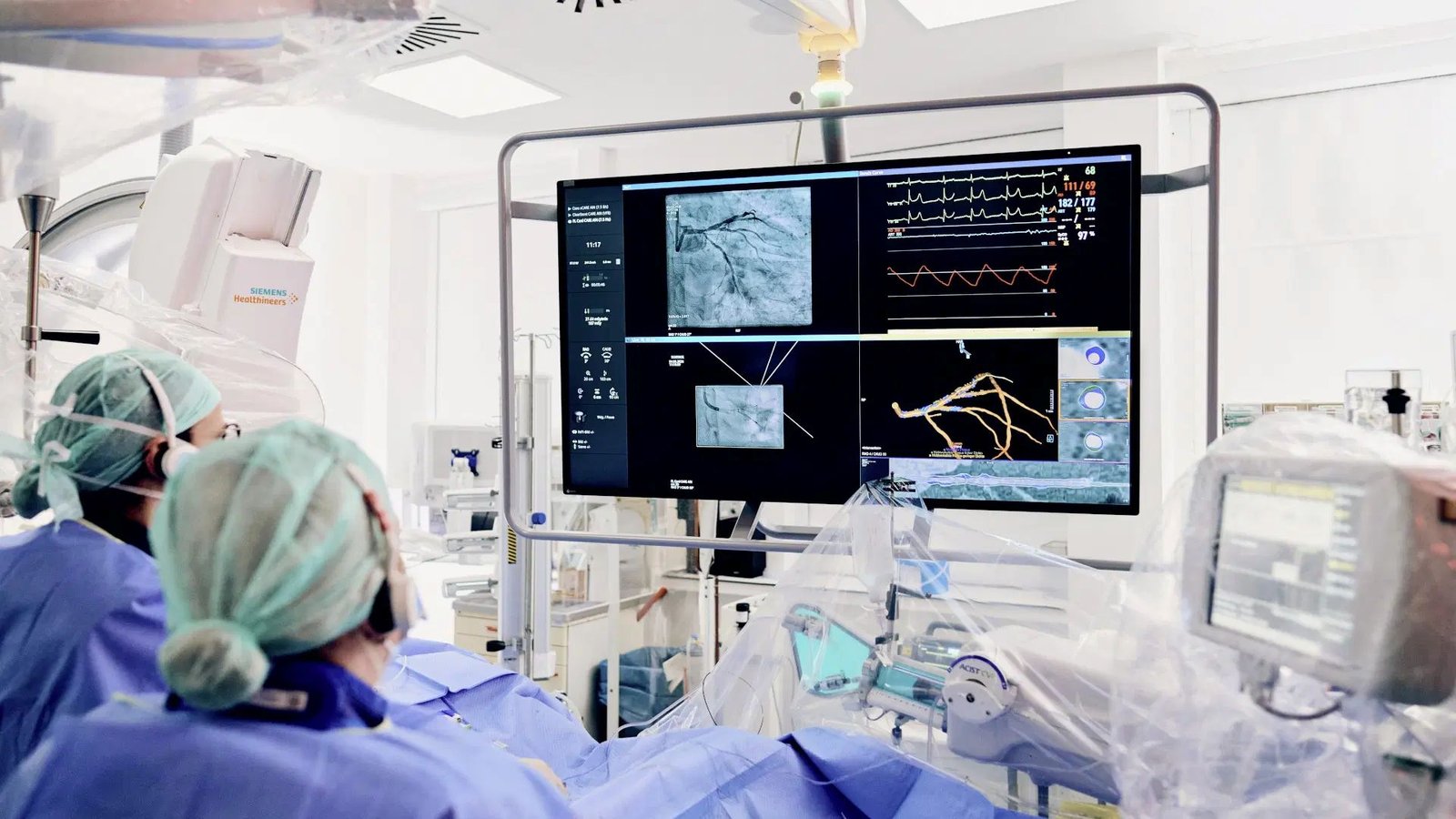
The Siemens Healthineers solution covers the complete CT-guided PCI workflow through the integration of Syngo.CT coronary Cockpit on the Syngo.via platform and Syngo PCI Connect on the new Artis angiography systems. Based on the CT data, the anatomical overview, plaque information and lumen dimension can be displayed in the cath lab, as well as side-by-side views and markers for planned stents.
The intuitive visualisation on the large display of the Artis system gives guidance during interventions and has all relevant information at hand. The C-arm and the CT images are automatically synchronised, which means that when the C-arm is moved, the CT image follows. A unique feature is that this synchronisation works both ways. When the interventional cardiologist adjusts the CT image to have the best possible view of the vessel, that precise angulation is sent to the C-arm, which moves there with a deflection of the joystick.
“Now that we can perform CT-guided PCI, we can integrate valuable information from CT into our angio system in the cath lab”, said Professor Samuel Tobias Sossalla, MD, director of cardiology at Kerckhoff Clinic Bad Nauheim and University Clinic Gießen, Germany. “We can define our PCI strategy upfront and plan our resources much better. During the procedure, we see this information synchronised with the corresponding angulations, helping us treat with greater precision.”