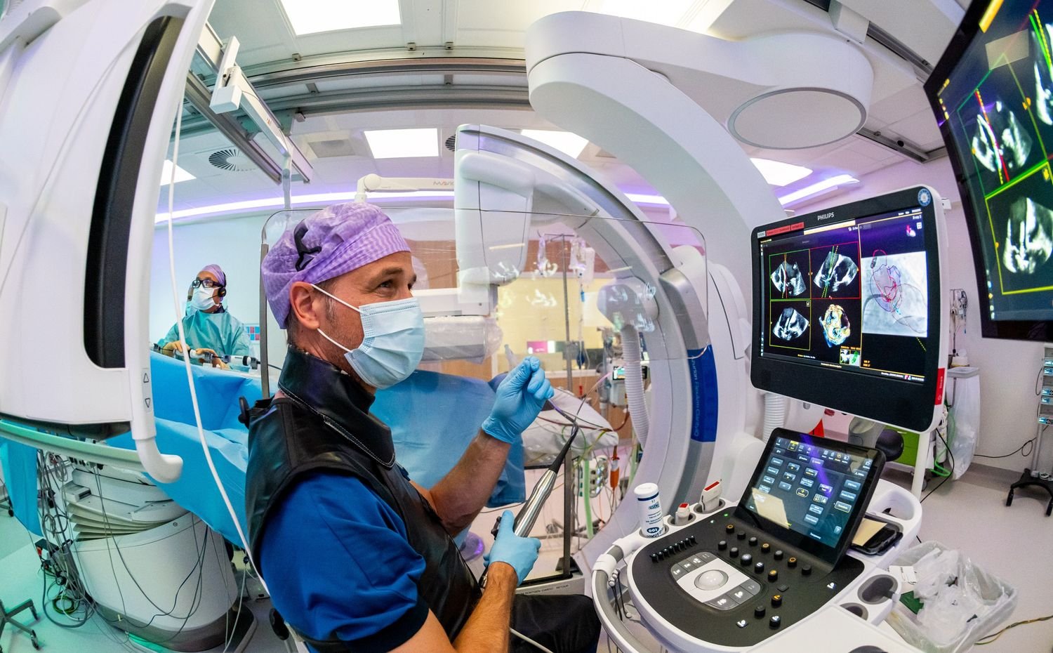
As healthcare systems worldwide grapple with ageing populations and the rising burden of chronic cardiovascular disease, the need for continuous, patient-centric monitoring has never been greater. While ultrasound remains a gold standard for cardiovascular imaging, its use has traditionally been limited to episodic, hospital-based examinations, leaving significant gaps in long-term disease monitoring and early intervention.
Addressing this challenge is the Wearable Imaging for Transforming Elderly Care (WITEC) initiative, led by researchers from the Singapore–MIT Alliance for Research & Technology (SMART). The programme aims to develop the world’s first wearable ultrasound imaging system capable of 48-hour continuous cardiovascular monitoring, enabling real-time insights into cardiac function beyond the confines of the hospital.
In this interview with Medtech Spectrum, Prof Xuanhe Zhao, Co-Lead Principal Investigator of WITEC and a faculty member at MIT, discusses the scientific and clinical drivers behind the initiative, the breakthroughs in materials science, microfabrication and AI that are making continuous ultrasound imaging possible, and the role of cross-institutional collaboration involving MIT, NTU, NUS and Tan Tock Seng Hospital. He also shares how this technology could accelerate the shift toward preventive, home-based healthcare and transform the management of chronic cardiovascular conditions.
WITEC aims to create the world’s first wearable ultrasound imaging system capable of 48-hour cardiovascular monitoring. What were the scientific or clinical gaps that motivated the inception of this initiative?
Ultrasound monitoring is largely episodic and hospital-based as traditional ultrasound systems are bulky and operator-dependent, and only provide snapshots – making them unsuitable for continuous, long-term, everyday use. Existing wearable devices, such as fitness trackers, offer only limited physiological data (e.g., heart rate, ECG) and cannot provide continuous imaging or monitor chronic conditions, especially during dynamic body motion.
There is a clear clinical need for continuous, real-time monitoring for earlier detection of symptom deterioration and timely intervention, particularly for chronic conditions like hypertension and heart failure.
Additionally, sonographers require extensive training to become proficient in cardiography. Patients also often remain in the hospital while awaiting their ultrasound examinations. By implementing long-term ultrasonic imaging, we can reduce the hospital's manpower and cost burdens, shifting the focus of healthcare from hospitals to patients' homes.
Wearable ultrasound represents a major shift from episodic to continuous monitoring. What breakthroughs in materials science, imaging technology, or data processing have made this vision technically possible today?
The project leverages bioadhesive technology for skin-safe, long-term adhesion and uses rigid ultrasonic transducers paired with a soft gel interface to ensure deep, consistent imaging quality even during movement.
Microfabricated Ultrasonic Transducer (MUT) technologies and Application-Specific Integrated Circuit (ASIC) technologies enable the monolithic integration and miniaturisation of transducers. Advanced semiconductor fabrication processes allow for high-quality, cost-effective and scalable device production.
WITEC’s lab is equipped with advanced tools, including the Verasonics Vantage NXT 256 ultrasonic imaging system – the first in Singapore – that enables complex beamforming and high-resolution image capture, and Southeast Asia’s first Nanoscribe Quantum X sub-micrometre 3D printer that allows researchers to fabricate components with sub‑micrometre resolution.
Finally, robust AI algorithms are needed to process the large volumes of data generated, ensuring accurate, real-time diagnostics without overwhelming clinicians or patients. These algorithms can enhance images and learn visual cues from extensive training data. They are capable of making diagnostic decisions even with image qualities that are lower than what is perceptible to the human eye. Additionally, these algorithms facilitate edge data processing, allowing data to be processed immediately after acquisition by the sensors. This is a departure from traditional designs, which require the complete set of raw data to be transmitted to the control unit before signal processing and image reconstruction occur. As a result, this approach significantly reduces the need for wiring and communication hardware.
The project brings together MIT, NTU, NUS, and TTSH. How does this interdisciplinary and multinational collaboration shape the research strategy, and what unique strengths does each partner contribute?
WITEC has assembled a diversified team of experts from MIT, NTU, NUS and TTSH – each bringing the best expertise in their respective fields to accelerate the translation of foundational research into clinical solutions.
MIT brings in its valuable expertise in soft materials, metamaterials and AI techniques. NTU excels in medical imaging, will oversee the clinical study while collaborating on the development of soft coupling materials. NUS, recognised for its strengths in medical devices and their translational applications, will provide essential guidance for integrating these innovations into clinical practice.
TTSH will lead a clinical study involving 50 patients admitted for acute decompensated heart failure and cardiogenic shock, validating the feasibility and safety of continuous ultrasound monitoring in an in-patient setting.
The Nanoscribe Quantum X 3D printer and Verasonics Vantage NXT 256 system provide capabilities not previously available in Southeast Asia. How will this advanced infrastructure accelerate device development and testing?
The Quantum X 3D printer enables the fabrication of metamaterials with submicrometre feature sizes; this size level has not been thoroughly explored, presenting new opportunities for controlling ultrasonic waves in the 1-30 MHz range. The Verasonics Vantage NXT 256 supports advanced imaging protocols and integration with AI models. It offers more channels than its predecessors and includes advanced features such as arbitrary waveform synthesis. These improvements open up new possibilities for larger ultrasonic array beamforming.
Long-term, skin-adherent ultrasound devices pose engineering and human-factor challenges. What are the key hurdles your team must overcome to ensure comfort, accuracy, and safety for 48-hour monitoring?
One of the foremost hurdles is designing bioadhesive materials that can securely attach the device to the skin for up to 48 hours, while maintaining comfort, minimising irritation and ensuring stable acoustic coupling. The WITEC team is innovating in soft materials to achieve low acoustic attenuation, skin-matching rigidity, high fracture toughness and strong yet comfortable adhesion.
To ensure consistent imaging quality during dynamic patient movement; WITEC’s approach uses rigid, high-resolution ultrasound probes with a soft gel interface, which avoids the imaging errors associated with stretchable or flexible transducers.
The device must also be compact and unobtrusive, so as not to interfere with daily activities, and must operate at low power to ensure safety — ultrasound is inherently non-ionising and safe for prolonged use.
If WITEC succeeds, how do you envision wearable cardiovascular imaging transforming chronic disease management and the broader shift toward preventative, home-based healthcare?
By shifting care from episodic, hospital-based interventions to proactive, home-based monitoring, we’re able to enable earlier detection of disease complications through continuous, real-time imaging — such as fluid accumulation in lungs that can depict heart failure — and provide timely feedback for both patients and clinicians.
This could reduce hospital admissions, shorten lengths of stay and minimise the need for expensive diagnostic procedures like CT or MRI scans. The vast dataset generated by wearable imaging would also support the development of AI-driven personalised diagnostics, empowering patients to manage their health more effectively and enabling clinicians to intervene before conditions worsen.