Enspectra Health announced the publication of pivotal clinical trial results in JAMA Dermatology, demonstrating that its handheld cross-modal imaging system enables physicians to accurately identify key histologic features of skin noninvasively. The study, known as the VISTA Trial, was conducted at multiple U.S. dermatology centres and evaluated the system's ability to visualise skin features typically seen only through invasive biopsy and histopathology.
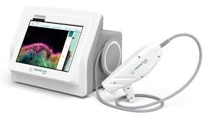
The cross-modal imaging system, VIO, combines reflectance confocal microscopy (RCM) and multiphoton microscopy (MPM) into a compact, handheld device that generates cross-sectional, histology-like images of the skin. In the trial, physicians trained in dermatopathology achieved 96.4 per cent accuracy in identifying primary histologic features and 98.5 per cent accuracy for secondary features, with high inter-reader agreement.
"This study shows that physicians can reliably identify histologic features such as collagen, pigment, hyperkeratosis, atypia, and solar elastosis using VIO's cross-modal imaging," said Dr Sarah Arron, lead author of the study. "These features are key to understanding skin health, and noninvasive visualisation opens new possibilities for both medical and aesthetic applications."
The trial enrolled 65 participants undergoing routine skin biopsy, with cross-modal imaging performed before tissue collection. All histologic features identified on cross-modal images were validated against ground truth pathology. The study population included 13.8 per cent Hispanic participants and Fitzpatrick skin types I–V, supporting the system's utility across a range of skin tones.
"During the trial, we imaged a range of common skin lesions across multiple anatomic locations and Fitzpatrick skin types I–V," said Dr Michael Wang, senior author and principal investigator. "This design allowed us to evaluate the VIO system's ability to capture histologic features in a variety of commonly encountered clinical contexts."