Artificial intelligence in healthcare is often discussed through algorithms, model performance and predictions about what hospitals may look like in the future. At Philips Pulse Connect in the Netherlands, the more revealing question was much more practical: what does AI actually do during a patient’s care journey?
Across two days at Philips’ innovation campus in Best and St. Antonius Hospital in Nieuwegein, the answer emerged through a series of demonstrations spanning diagnostic imaging, cardiac monitoring, maternal ultrasound and image-guided heart treatment.
Rather than presenting AI as a separate layer sitting above clinical care, Philips showed technologies designed to operate within the individual steps that clinicians already perform: positioning a patient, planning a scan, reconstructing an image, reviewing a long ECG recording or navigating a device inside a beating heart.
The common objective was not simply to make individual systems more advanced. It was to reduce the manual work, variability and dependence on scarce expertise that limit how many patients can receive timely diagnosis and treatment.
That distinction matters particularly across Asia-Pacific, where rising imaging volumes, ageing populations and uneven access to specialists place pressure on healthcare systems at both ends of the spectrum—from high-throughput urban hospitals to rural clinics with limited diagnostic infrastructure.
From an AI presentation to a working scanner
The first day presented us with the wider healthcare challenge and provided context to the problems and solutions that would be presented to us.
Patrick Mans, Global Head of Data and AI at Philips, pointed to the projected global shortage of around 10 million healthcare professionals by 2030. He presented that healthcare systems cannot recruit their way out of the problem.
AI, in this case, is less about replacing the clinical workforce than absorbing parts of the workload that keep trained professionals away from patients. These include documentation, administrative tasks, equipment setup, image preparation and the review of large volumes of routine clinical data.
That argument became more tangible when we moved from the conference room into Philips’ demonstration spaces.
At the CT station, the scale of the operational challenge quickly became clear. A scanner in a typical Dutch department may examine about 30 to 50 patients during a working day. In high-volume markets across Asia-Pacific and South America, the same type of system may need to handle 150 to 250 patients.
A CT examination itself can be completed in seconds, but the surrounding workflow still includes patient positioning, protocol selection, scan planning, reconstruction and post-processing. Every manual adjustment consumes time and introduces another opportunity for variation between operators.
Philips is embedding AI across this workflow, including automatic patient positioning and image reconstruction. The practical aim is to enable a scanner to move more consistently from one patient to the next while maintaining image quality and managing radiation exposure.
Standing beside the system, the value of automation was shown clearly. At high patient volumes, saving a few interactions and clicks on one scan may appear minor. Considering repeated across hundreds of examinations, however, those interactions become hours of staff capacity.
Towards a ‘self-driving’ MRI
The cardiac MR demonstration addressed a different problem.
Cardiac magnetic resonance imaging can provide detailed information on the structure and function of the heart and support the assessment of conditions including heart failure, cardiomyopathies, inflammation and scarring. Yet the examination remains complex, lengthy and dependent on trained personnel.
Philips estimates that around half of the patients who could benefit from cardiac MR do not receive the examination in a timely manner.
The demonstration used the analogy of a self-driving car to explain the company’s direction for MRI. The scanner is not being designed to remove the technologist or radiologist from the process. Instead, sensors, cameras and software progressively take over repeatable elements while the clinician remains responsible for the patient and the examination.
One example was an AI-powered cardiac MR planning solution designed to automate the planning process of cardiac MRI exams, which automatically identifies cardiac anatomy and creates the required imaging views. Philips said the technology can reduce scan-planning interactions from approximately 128 clicks to three.
An AI-powered MR software solution then applies AI during image acquisition and reconstruction to accelerate the scan while preserving diagnostic image quality.
The significance is not only a faster examination. Simplifying cardiac MR could allow the procedure to move beyond highly specialised centres and become available in more hospitals, including those without large teams of cardiac imaging experts.
Finding the signal in days of heart rhythm data
At another station, a small wearable patch provided a very different illustration of the same principle.
This solution is used for extended cardiac monitoring and can be worn for seven or 14 days, compared with the 24- or 48-hour monitoring periods commonly associated with conventional Holter systems.
Longer monitoring increases the likelihood of capturing intermittent arrhythmias such as atrial fibrillation. The patient can shower, perform light exercise and continue much of their daily routine, making it more likely that the conditions that trigger an irregular rhythm will occur during the monitoring period.
However, several days of continuous ECG recordings generate far more information than a clinician could efficiently review heartbeat by heartbeat. To counter this, Philips’ ECG analysis solution applies AI to analyse the recording and highlight clinically significant events, leaving the cardiologist or technician to review the sections that may require action.
This was one of the clearest examples of AI supporting rather than making the final clinical decision. The system filters and prioritises, while the clinician interprets the result and determines the diagnosis and treatment.
The operational implications can be significant. One Dutch hospital increased its annual volume from around 2,000 Holter studies to approximately 4,000 wearable studies without increasing staff numbers, according to Philips.
AI guidance outside specialist hospitals
The most compact technology shown during the visit may have the broadest access implications.
An AI-guided obstetric ultrasound application was being developed for use by nurses and midwives in lower-resource primary care settings.
Traditional obstetric ultrasound depends on both equipment and a trained operator who can acquire and interpret the required images. In remote communities, the absence of a sonographer can make the equipment itself of limited use.
The application takes a different approach. A healthcare worker moves a portable ultrasound probe across the patient’s abdomen while the application provides real-time audio-visual guidance and identifies scanning errors. The system generates automated obstetric measurements without requiring the operator to interpret conventional ultrasound images.
The demonstration therefore moved the emphasis from producing an image to producing the information needed to identify whether a pregnancy may require referral.
This does not remove the need for maternity services, referral pathways or higher-level clinical expertise. It may, however, enable primary care workers to identify higher-risk pregnancies earlier and direct patients to appropriate care.
For Asia-Pacific, where specialist availability and travel distances vary considerably between and within countries, this model may be as relevant as the advanced imaging technologies used in large hospitals.
From diagnosis to treatment
The most compelling part of the programme came when the discussion moved from diagnosing disease to treating it.
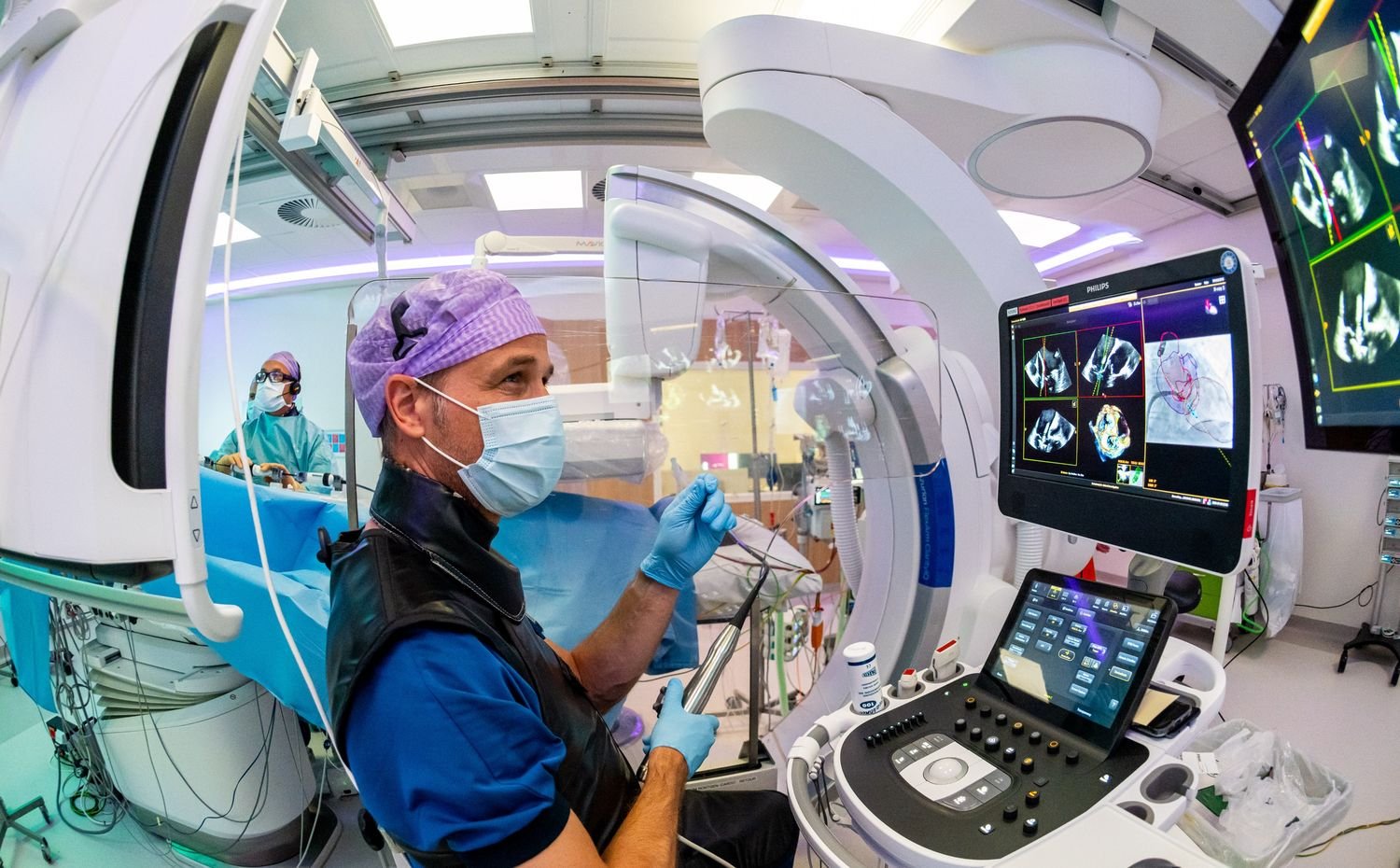
At Philips’ hybrid operating room demonstration, The AI-based real-time guidance solution supports structural heart procedures was introduced as an AI-enabled application for structural heart procedures. The technology combines echocardiography and X-ray imaging to help physicians visualise both the patient’s anatomy and the treatment device.
In conventional open-heart surgery, a surgeon can directly see the valve being repaired. During a minimally invasive transcatheter procedure, there is no direct line of sight. The clinical team depends on imaging to navigate through the heart, position the device and assess the result.
The AI-based guidance solution identifies the treatment device within echocardiography images and creates a visual representation that can be fused with live X-ray. The intention is to give the interventional cardiologist a clearer understanding of the device’s position and orientation inside a complex, moving structure.
The next day, that explanation moved into a real hospital environment.
Seeing intelligent treatment in the Cath Lab
At St. Antonius Hospital, cardiologists Dr Martin Swaans and Dr Leo Timmers introduced the clinical realities of treating mitral regurgitation, a condition in which the mitral valve does not close properly and allows blood to flow backwards through the heart.
The mitral valve is structurally complex, and many patients considered for transcatheter treatment are older or may not be suitable candidates for open surgery.
Before entering the Cath laboratory, the two physicians described their roles simply: one acts as the navigator, while the other controls the treatment device. That description became clearer once the procedure began.
From outside the sterile area, we could follow the intervention through multiple live imaging views. Echocardiography showed the valve and surrounding soft tissue. X-ray showed the catheter and treatment device. The AI-based real-time guidance solution brought those sources together, allowing the team to see how the device was approaching the valve.
The procedure was neither silent nor automated. It depended on constant discussion between the physicians, imaging specialists, nurses and other members of the team. AI did not replace that interaction. It created a shared visual reference around which the team could communicate.
Unlike open surgery, the patient’s heart continued beating throughout the intervention. This allowed the physicians to assess the effect of the repair immediately. They could see whether regurgitation had been reduced and make adjustments before completing the procedure.
Watching the team work made the idea of an AI “co-pilot” more credible than any presentation slide could. The technology did not take control of the procedure. It helped the clinical team understand where the device was and what to do next.
The gap between availability and adoption
The visit also made clear that technical capability alone will not determine whether healthcare AI improves access.
During the plenary session, we enquired what infrastructure is required for AI to be implemented successfully across healthcare systems.
Carla Goulart Peron, Chief Medical Officer at Philips, commented that AI should not simply be inserted into an existing process. Hospitals and care pathways may need to be redesigned around how patients, data and clinical decisions move through the system.
That includes more than digital infrastructure. It involves referral networks, workforce training, reimbursement, governance and the ability to connect different parts of a patient’s care.
This was particularly relevant when comparing the technologies shown during the event.
A highly automated MR scanner may expand imaging capacity, but patients still need access to the scanner and a clinician who can act on the findings. The AI-guided obstetric ultrasound application may allow a nurse to identify a high-risk pregnancy, but its value depends on whether the patient can be referred to an appropriate facility. Remote support may help clinicians perform advanced procedures, but hospitals still need trained teams and suitable treatment infrastructure.
What the experience showed
The most important impression from Philips Pulse Connect was not that AI is becoming more powerful. It was that the technology is becoming less visible.
In the examples shown, AI was embedded in equipment controls, scan planning, image reconstruction, clinical data review and procedural navigation. It worked behind the interfaces clinicians already use.
That may be the more realistic direction for healthcare AI adoption. Hospitals are unlikely to transform through a single algorithm or platform. Change will come through hundreds of smaller workflow decisions: fewer manual steps, more consistent imaging, earlier identification of risk and better access to specialist guidance.
At St. Antonius Hospital, the result still depended on the judgement and coordination of an experienced clinical team. Yet the team was able to see and navigate the procedure in ways that would not have been possible through conventional imaging alone.
The experience brought Philips’ wider proposition into focus. The next phase of healthcare AI may not be defined by machines replacing clinicians, but by technology helping clinical knowledge travel further. Integration would be key, as we advance alongside technology.