
Australian medical technology company OncoRes Medical has adopted the trading name Ziva Surgical as it advances its investigational ELORA tissue imaging system through clinical development and prepares for commercialisation.
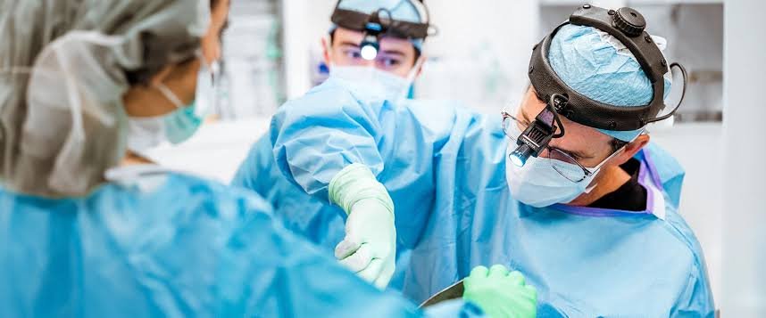
ELORA uses quantitative micro-elastography to generate microscale maps of tissue stiffness and structure. The system is intended to help surgeons assess tissue within the surgical cavity and decide what to remove or preserve during an operation.
Its initial application is breast-conserving surgery, where identifying residual cancer remains a challenge. The company estimates that approximately one in five women undergoing these procedures requires another operation to remove remaining cancerous tissue.
The technology emerged from research at the University of Western Australia, the Western Australian Department of Health and the Harry Perkins Institute of Medical Research. OncoRes was established as a university spinout in 2016, building on work led by breast cancer surgeon Professor Christobel Saunders and biomedical engineer Professor Brendan Kennedy.
Earlier in 2026, the company secured $27 million in private funding to advance clinical trials, product development, manufacturing and regulatory milestones. An Australian interventional study was designed to recruit 110 breast cancer patients across six hospitals in Western Australia and Victoria.
Chief Executive Officer Dr Katharine Giles said the company is approaching important clinical milestones, with results from its Australian trial expected soon.
ELORA received FDA Breakthrough Device Designation in 2020. The system remains investigational, and the designation does not constitute marketing authorisation.
Ziva Surgical is also exploring applications beyond breast surgery, including prostate cancer. Its imaging approach is designed to complement pathology by providing additional information during surgery, rather than replacing the examination of tissue specimens.