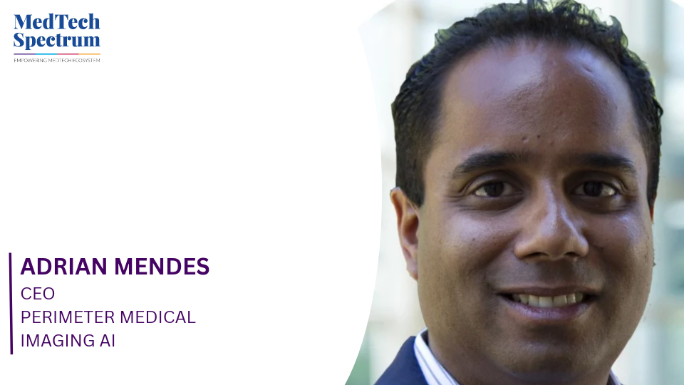
As breast-conserving surgery continues to play an important role in breast cancer treatment, identifying microscopic cancerous tissue at surgical margins remains a challenge, often leading to repeat procedures. Perimeter Medical Imaging AI is addressing this gap with Claire, an AI-enabled optical coherence tomography (OCT) imaging system designed to provide surgeons with real-time insights into excised breast tissue during surgery. In this interview with MedTech Spectrum, Adrian Mendes, CEO of Perimeter Medical Imaging AI, discusses how Claire combines high-resolution imaging and AI to support intraoperative margin assessment, the importance of clinical validation and workflow integration, and the technology’s potential to reduce re-excision rates. He also outlines the company’s priorities following FDA approval and its plans to explore applications in other cancer surgeries.
Could you explain how Claire uses optical coherence tomography (OCT) and AI to help surgeons identify microscopic cancerous tissue at the margins during breast-conserving surgery? What differentiates this technology from conventional intraoperative imaging methods?
Claire is the first FDA-approved, AI-enabled imaging device for intraoperative margin assessment during breast-conserving surgery in the U.S. Optical Coherence Tomography (OCT) is a light-based imaging technology that’s been trusted in ophthalmology and cardiology since the 1990s. We adapted it for soft tissue so it can be used adjunctively inside the operating room. Once the surgeon removes the specimen, Claire scans it up to 2 mm below the tissue surface and produces high-resolution 3D images of the tissue within minutes. Our proprietary AI, trained on more than 2 million pathologically correlated breast tissue images, highlights areas that may be suspicious for cancer to guide the surgeon where to take a closer look. The clinical decision always stays with the surgeon.
The difference from conventional tools comes down to resolution and what the surgeon can actually see. Specimen X-ray and ultrasound are useful for locating clips and larger structures, but they can’t identify microscopic disease at the tumor margin (the outer edge of the removed tissue, which is checked for cancer cells to assess whether the cancer was fully removed). Other approaches in this space rely on radiofrequency, which can signal that something may be present without giving the surgeon an image of the tissue itself. Claire's imaging is about 10x sharper than ultrasound or specimen X-ray and 100x more detailed than MRI. Because the Claire device scans the excised specimen, the device never touches the patient, requires no dyes, drugs, or ionizing radiation, and leaves the tissue fully intact for pathology.
Breast cancer re-excision remains a significant clinical challenge, with many patients requiring a second procedure due to post-operative detection of residual disease. How is Claire designed to transform decision-making in the operating room and potentially reduce re-excision rates?
Today, about 1 in 5 lumpectomy patients need a second surgery because microscopic cancer cells at the margin weren’t visible, and therefore weren’t removed, during the first procedure. For women ages 18 to 44, that number climbs to 40 per cent. The core issue is visibility: surgeons can't always see microscopic cancer at the tissue's edge during the procedure. Final pathology, which can take up to 7 days, is still needed to determine whether the margins are clear, even when Claire is used. If cancer remains, patients may have to come back in for another operation to remove the rest of the diseased tissue.
Claire is designed to bring additional information into the operating room during the window when the surgeon can still act. If the AI flags an area of concern, the surgeon can evaluate it and take additional tissue before the patient leaves the operating room. In our multicenter pivotal trial, Claire demonstrated 88.1 per cent margin accuracy and met its primary endpoint, showing a statistically significant reduction in patients with residual cancer when used alongside standard of care compared with standard of care alone. Additional tissue removed under Claire's guidance accounted for just 4 per cent of the average total breast tissue volume excised. Preserving healthy tissue matters because removing more can change the breast's appearance and be emotionally difficult for patients. No surgeon wants to remove more tissue than necessary.
The stakes go beyond the clinical picture. No surgeon wants to make the call telling a patient they need another surgery, and no patient wants to receive it. Repeat surgeries are linked to a 66% higher risk of post-operative complications, delayed treatment, and can have a psychological impact. When a patient requires a second surgery, their satisfaction with choosing a lumpectomy drops from 76 per cent down to 47 per cent, while negative emotional well-being doubles from 17 per cent to 35 per cent. Beyond the human toll, repeat surgeries cost the U.S. healthcare system an estimated $600 million each year. Giving surgeons better information during the first procedure has the potential to reduce how often patients have to come back.
Given your background in AI chip infrastructure at Groq, how has your experience building AI systems influenced your approach to developing clinical AI, particularly in ensuring technology integrates seamlessly into existing surgical workflows?
At Groq, I worked on AI from the silicon up, and I saw firsthand what it takes to move advanced technology out of the lab and into real-world use under pressure and at scale. One of the biggest lessons was that compute, speed, data architecture, and cost only create value when the tradeoffs are weighed against a specific problem that needs solving. Technical performance on its own doesn't carry a product. Usability within the clinical setting adds additional complexity that was demonstrated in support of Claire’s PMA approval.
That thinking applies directly to the operating room. Surgeons can't pause a procedure to interpret a separate data stream or wait on another system. If a tool adds steps or slows the team down, it won't get used, no matter how good the underlying technology is. So we aimed for Claire to exist within the surgeon's existing workflow by providing focused, actionable information in minutes. The AI acts as a guide that points attention to areas worth a second look, so the surgeon can decide in real time in the OR whether more tissue should be removed. My background also helps us think about how the full system, from data to algorithm, can keep improving over time.
What role does AI play in interpreting Claire's OCT images, and how have you addressed the challenges of clinical validation, accuracy, and reliability in a high-stakes surgical environment?
OCT provides a level of resolution that other modalities can't match and produces a tremendous amount of visual data for the surgeon to review. The AI helps streamline that review by flagging microstructures associated with ductal carcinoma in situ (DCIS), a noninvasive form of breast cancer confined to the milk ducts, and invasive ductal carcinoma (IDC), so surgeons can review the data faster and focus on the areas that matter most. Reliability starts with the data. Claire was trained on more than 2 million proprietary breast tissue images from approximately 4,000 real-world surgeries, all correlated with pathology and specific to this clinical use case.
During surgery, our models are locked to protect patient safety. Claire was also approved with a Predetermined Change Control Plan (PCCP), which gives us an FDA-reviewed framework for refining the algorithm over time as we gather more real-world clinical data. As more images are added, that framework could allow us to improve the AI's precision, potentially helping surgeons make more informed decisions and improving patient outcomes.
Beyond breast-conserving surgery, what future opportunities do you see for OCT-based imaging and AI in other surgical or diagnostic applications where real-time identification of microscopic disease is critical?
Positive margins are a challenge across many solid tumor surgeries, not only breast cancer. Because our OCT platform has shown potential to distinguish multiple tissue types, we’re planning clinical and regulatory work in other areas where clear margins are equally important, including head, neck, and lung cancer.
Over the longer term, we see potential for the platform for biopsy and pathology applications as well. Any of those uses would require their own clinical evidence and regulatory review, and our breast surgery work gives us a strong model for how to build that evidence.
Following Claire's FDA approval, what are the next priorities for Perimeter Medical Imaging regarding clinical adoption, evidence generation, healthcare system expansion, and patient accessibility?
Our near-term priority is expanding hospital adoption as we move from a regulatory-focused company to a commercial, customer-driven one. Intermountain Health in Salt Lake City became the first U.S. health system to deploy Claire, and we’re working to build on that momentum with additional health systems.
Evidence generation runs alongside adoption. Real-world use helps us build the clinical and economic case for Claire and continues to strengthen the dataset behind the AI, which we can refine through our FDA-reviewed change control pathway.
Accessibility is also important to us. Surgeons who perform fewer breast cancer procedures could gain access to the kind of real-time decision support often associated with high-volume centers, which may help improve consistency of care across settings. Our long-term goal is for real-time margin assessment to become the standard of care in breast-conserving surgery, so more patients may only have to endure one procedure to fully remove all cancer.
References
Disclaimers
The Claire OCT System is an adjunctive three-dimensional imaging tool which provides volumetric cross-sectional, real-time depth visualization, coupled with an artificial intelligence computer-aided detection algorithm that identifies and marks focal areas suspicious for breast cancer and is used concurrently with physician interpretation of the images. Claire is intended for use in conjunction with other standard methods for evaluation of the margins of excised lumpectomy tissue during surgical procedures in patients with a biopsy-confirmed diagnosis of breast cancer. Perimeter’s OCT technology has not been evaluated by the FDA for use in any other tissue nor health condition. Claire should not be used to replace standard tissue histopathology assessment nor used for diagnosis. The efficacy of Claire was evaluated only after completion of standard of care (SOC), in accordance with the pivotal trial design supporting PMA P250008. For additional details regarding the study design and clinical effectiveness results, please refer to https://clinicaltrials.gov/study/NCT05113927.